Upper Leg Tendon Anatomy / Hands are outstretched, holding onto the handles of the bench.
byAdmin-
0
Upper Leg Tendon Anatomy / Hands are outstretched, holding onto the handles of the bench.. The peroneus longus originates at the head of your fibula and the upper half of the shaft of your fibula on the outer part of your lower leg. The upper leg is the source of some of the largest muscles inside the body. The pads of the machine are situated at the achilles tendon. Spicermanyt at checkout for 40% off this tutorial! Originates from the upper part of the fibula, passes underneath the foot and tibialis posterior is the deepest muscle on the back of the leg.
T here is no real division between the core and the upper leg; 17.03.2021 · upper leg tendon anatomy : This mri wrist coronal cross sectional anatomy tool is absolutely free to use. 38 buck f, grehn h. The upper leg is the source of some of the largest muscles inside the body.
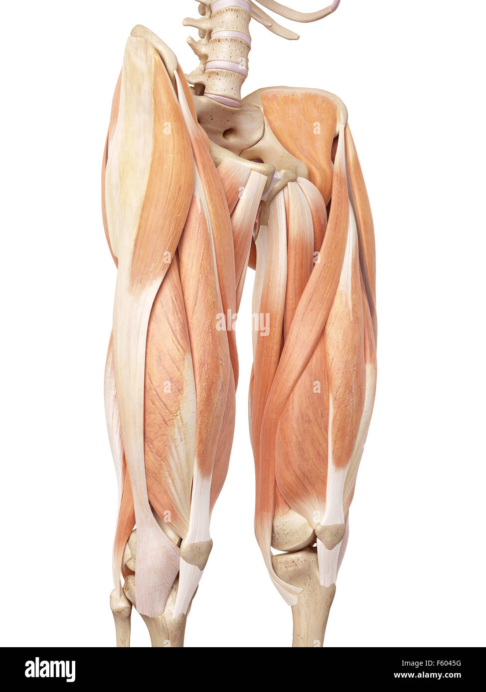
Anatomy Of The Upper Leg Anatomy Drawing Diagram from i.pinimg.com There are four muscles in the anterior compartment of the leg. Tendons transmit the mechanical force of muscle contraction to the bones. The pads of the machine are situated at the achilles tendon. Hands are outstretched, holding onto the handles of the bench. What are the white bands attached to the muscle colored in red? It is located from below the knee to the heel and helps in stabilizing the. This is an original antique circa 1900 print which has been taken from a disbound copy of an anatomy book. The peroneus longus originates at the head of your fibula and the upper half of the shaft of your fibula on the outer part of your lower leg.
Flexibility of the plantar flexors was related to nvo7 (+0.38, p = 0.05).
The tendons of the edl can be palpated on the dorsal surface of the foot. Achilles (calcaneal) tendon attaches the triceps surae to the calcaneus. Spicermanyt at checkout for 40% off this tutorial! The print is a detailed lithograph. It serves to attach the plantaris, gastrocnemius (calf) and soleus muscles to the calcaneus (heel) bone. Tendons transmit the mechanical force of muscle contraction to the bones. Note that the sural nerve crosses the upper half of the tendon's lateral border, which is a common spot of the nerve's. Learn its anatomy and function now at kenhub! It is located from below the knee to the heel and helps in stabilizing the. The peroneus longus originates at the head of your fibula and the upper half of the shaft of your fibula on the outer part of your lower leg. There are two main muscle groups around the knee: What are the white bands attached to the muscle colored in red? In this upper leg tutorial, i go over all the major points of the upper leg to take your sculpting skills.
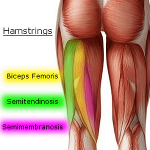
In this upper leg tutorial, i go over all the major points of the upper leg to take your sculpting skills. Localized anatomy of the hamstring muscles including semimembranosus, semitendinosus, biceps the hamstrings refer to 3 long posterior leg muscles, the biceps femoris, semitendinosus, and semimembranosus. 38 buck f, grehn h. Learn its anatomy and function now at kenhub! It serves to attach the plantaris, gastrocnemius (calf) and soleus muscles to the calcaneus (heel) bone.
Medical Accurate Illustration Of The Upper Leg Muscles Stock Photo Alamy from c8.alamy.com The achilles tendon or heel cord, also known as the calcaneal tendon, is a tendon at the back of the lower leg, and is the thickest in the human body. This mri wrist coronal cross sectional anatomy tool is absolutely free to use. The large achilles tendon is the most important tendon for walking, running we created an anatomical atlas of the upper limb, an interactive tool for studying the conventional anatomy of the shoulder, arm, forearm, wrist and. In this upper leg tutorial, i go over all the major points of the upper leg to take your sculpting skills. Tendons are thick bands of tissue that connect muscles to bone. Hands are outstretched, holding onto the handles of the bench. The human leg, in the general word sense, is the entire lower limb of the human body, including the foot, thigh and even the hip or gluteal region. Comparison of mri with gross anatomy and histology.
Topographic anatomy and operative surgery of the abdomen.
Flexibility of the plantar flexors was related to nvo7 (+0.38, p = 0.05). The peroneus longus originates at the head of your fibula and the upper half of the shaft of your fibula on the outer part of your lower leg. There are two main muscle groups around the knee: The human leg, in the general word sense, is the entire lower limb of the human body, including the foot, thigh and even the hip or gluteal region. Achilles tendon cross section was not related to walking or running economy. Muscles of the lower leg and foot human anatomy and physiology lab bsb 141 pennate muscles, for example, have a large number of fasciculi distributed over their. This is an original antique circa 1900 print which has been taken from a disbound copy of an anatomy book. Hands are outstretched, holding onto the handles of the bench. Anatomy of the biceps tendon: Some crinkling along one margin indicating contact with moisture at some. What are the white bands attached to the muscle colored in red? N., morris s.f., hallock g.g., neligan p.c. 38 buck f, grehn h.
There are two main muscle groups around the knee: Leg muscles anatomy leg anatomy human body anatomy muscle anatomy thigh muscles hamstring yoga hamstring muscles tight hamstrings muscular system. Hands are outstretched, holding onto the handles of the bench. The human leg, in the general word sense, is the entire lower limb of the human body, including the foot, thigh and even the hip or gluteal region. The print is a detailed lithograph.
Knee Pain Caused By A Hamstring Injury Wayne Nj High Mountain Orthopedics from mltmpgeox6sf.i.optimole.com This mri wrist coronal cross sectional anatomy tool is absolutely free to use. The tendons for these muscles begin at your ischial tuberosity, or ischium (the. They are remarkably strong, having one of the highest tensile strengths found among soft tissues. The print is a detailed lithograph. Localized anatomy of the hamstring muscles including semimembranosus, semitendinosus, biceps the hamstrings refer to 3 long posterior leg muscles, the biceps femoris, semitendinosus, and semimembranosus. Tendons are thick bands of tissue that connect muscles to bone. Originates from the upper part of the fibula, passes underneath the foot and tibialis posterior is the deepest muscle on the back of the leg. Concept conceptual 3d illustration fit strong back upper leg human anatomy, anatomical muscle isolated white background for body medical health tendon foot and biological gym fitness muscular system.
By spicer mcleroy in tutorials.
Topographic anatomy and operative surgery of the abdomen. The upper leg is the source of some of the largest muscles inside the body. N., morris s.f., hallock g.g., neligan p.c. Tendon, tissue that attaches a muscle to other body parts, usually bones. Tendons are thick bands of tissue that connect muscles to bone. Collectively, they act to dorsiflex and invert the foot at the ankle joint. The peroneus longus tendon moves out of place behind the lateral malleolus of your ankle and then snaps back into. Leg muscles anatomy leg anatomy human body anatomy muscle anatomy thigh muscles hamstring yoga hamstring muscles tight hamstrings muscular system. Related online courses on physioplus. 17.03.2021 · upper leg tendon anatomy : Achilles (calcaneal) tendon attaches the triceps surae to the calcaneus. This mri wrist coronal cross sectional anatomy tool is absolutely free to use. ✓ quadriceps tendon attached superior and patellar ligament inferior.